An unobstructed view into the cell
Using a combination of the latest technological developments in the area of cryo-electron tomography, scientists from the Max Planck Institute of Biochemistry in Martinsried captured three-dimensional images of previously concealed structures in and around the nucleus of HeLa cells. They now present for the first time images of the nuclear lamina, a nanometer-thin filamentous protein structure that supports the nucleus, in the journal Science. The cellular components are observed in their natural environment without the cells being chemically altered or dehydrated. This is the only approach that allows visualization and understanding of the interactions between different functional components of a cell.

Thanks to the latest technological developments, scientists are able to penetrate into ever-smaller worlds. The aim of the research group led by Wolfgang Baumeister, Head of the Department of Molecular Structural Biology at the Max Planck Institute of Biochemistry, is to make the smallest cellular structures visible in their natural environment. As pioneers in the development of cryo-electron tomography, the researchers are now able to produce three-dimensional images of molecules like DNA and individual protein complexes and analyse them inside cells.
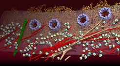
“Ninety percent of a cell consists of water. For traditional electron microscopy approaches, the cells are fixed, dehydrated, stained and, therefore, severely altered. This gives rise to artefacts and the functional context of the individual cellular components is lost,” explains Julia Mahamid, first author of the publication. “Sample preparation and imaging technologies have improved so much over the last five years that we can now see the four-nanometer-thick nuclear lamina for the first time,” she continues. This layer consists of thin thread-like proteins and supports the structure of the nucleus in cells. The 3D images also enabled the scientists to see nuclear pores, portals from the nucleus to the cytoplasm, ribosomes – the cell’s protein factories – and parts of the cellular skeleton, e.g. microtubules, intermediate filaments and actin.
The cells are snap-frozen for cryo-electron tomography. This makes the water in the cell freeze in a glass-like state. Ice crystals, which would destroy molecular structures, do not form. Transmission electron microscopy requires very thin samples. “Using a focused ion beam, approximately 200 nm-thin sections are produced in the snap-frozen human cells, which are typically a few micrometers thick,” as Mahamid explains the method, which was mainly employed in materials science up to now. Unlike the mechanical sections produced with the help of traditional methods, the molecular structures in the glass-like sections produced by this procedure remain unaltered.
Similar to computer tomography in medical diagnostics, two-dimensional images are recorded from the frozen cell thin sections at various angles using a transmission electron microscope. With the help of a computer, a three-dimensional image of the cellular landscape is then reconstructed from the recorded images. The Volta phase plate, which was recently developed at the institute, enables the scientists to obtain considerably enhanced contrast and provides unobstructed views into the densely packed nucleus.
“This project is a result of the combination of different innovations, which became possible in recent years, and is based on the joint contributions of many scientists,” says Mahamid. “In the future, it will be possible to make many cellular processes visible at molecular level in the natural environment.”
Originalpublikation:
J. Mahamid, S. Pfeffer, M. Schaffer, E. Villa, R. Danev, L. Kuhn-Cuellar, F. Förster, A. A. Hyman, J. M. Plitzko, W. Baumeister: Visualizing the molecular sociology at the HeLa cell nuclear periphery, Science, Februar 2016
DOI: 10.1126/science.aad8857


