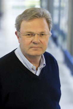
Wolfgang Baumeister wins Stifterverbandspreis 2019
The biophysicist has revolutionized molecular structural biology with the help of cryo-electron tomography.

Wolfgang Baumeister is to be recognized for his outstanding achievements in the field of cryo-electron tomography. What is particularly notable here is his examination of molecular and supramolecular structures within the context of intact cells at high spatial resolutions. The jury explained that it was awarding the prize because of the method’s great economic relevance, which is evident in important areas of high technology, such as electronics, materials technology and pharmaceuticals. The cryo method allows larger spatial structures from cells to electronic components to be tomographically captured at very high resolutions and analyzed.
Pioneering work in the field of cryo-electron tomography
“We are developing methods to make the molecular architecture of cells visible,” said Wolfgang Baumeister summarizing his research focus. The cryo-electron tomography method that he has developed in conjunction with his team opens up completely new opportunities for structural research: Entire cells or cell organelles are ‘shock-frozen’ in liquid nitrogen in the blink of an eye. The fragile cell architecture remains unchanged while it is embedded in the glassy ice. Two-dimensional images of the samples to be examined are taken from different angles. A three-dimensional image is then built from these images. This method has already made it possible to understand the architecture of many proteins in their cellular environments.
Structural research using electron microscopes
“It is only possible to understand the various functions that the molecular machines are responsible for by examining their structures,” explained Baumeister. Using cryo-electron tomography, he and his team have been able to decipher the structure of the 26S proteasomes, which are highly complex molecular shredders for proteins that are made up of 66 individual proteins. He and his team have also been able to reveal the superior organization of the ribosomes in cells, the so-called polysomes. The researchers have now turned their attention to other cellular structures and are investigating the blueprints of pores in nuclear membranes, contact points between nerve cells (synapses) and protein complexes in membranes and cell walls. It is possible to use cryo-electron tomography to image these macromolecular structures in the intact cellular environment. Pathological changes, such as toxic protein aggregates that are mainly associated with such neurodegenerative diseases as Alzheimer's and Parkinson's, may also be investigated in this way. This method that has been developed in basic research is potentially able to open up new prospects for therapies by providing insights into cell architectures.


