New synapse type discovered through spatial proteomics
Researchers led by Ralf Jungmann developed a super-resolution imaging method (SUM-PAINT) to map protein distributions in neurons and discovered a new type of synapse. SUM-PAINT makes it possible to study the diversity of synapses and explore the complexity of neurological diseases.
Researchers led by Ralf Jungmann at the Max Planck Institute (MPI) of Biochemistry and Ludwig-Maximilians-Universität (LMU) Munich, in collaboration with Eugenio F. Fornasiero, group leader at the Department of Neuro- and Sensory Physiology at the University Medical Center Göttingen (UMG), Felipe Opazo, group leader at the Department of Neuro- and Sensory Physiology and at the Center for Biostructural Imaging of Neurodegeneration (BIN) at UMG, and Helmholtz Munich, have developed a new super-resolution high-throughput imaging method. Using the new technique, the scientists were able to create a 3D neuronal cell atlas with single-molecule resolution and discovered a previously unknown type of synapse. The results of the study were published in the journal Cell.
In the current study, led by Eduard Unterauer in Jungmann's laboratory at the MPI of Biochemistry and the LMU, the researchers present SUM-PAINT. This is a new technological development in super-resolution microscopy that, for the first time, allows very fast and virtually unlimited visualization and mapping of a large number of proteins.
"The complexity of living systems ranges from entire organisms and tissues, to the structure of intricate cellular networks, to the organization and interaction of individual biomolecules. To understand this complexity in its entirety, the position, identity, and interaction of individual biomolecules must be studied simultaneously. Such methods, which combine multiple signals, are called multiplexing methods. Four critical challenges must be overcome to achieve a comprehensive understanding of protein organization: Sensitivity, throughput, spatial resolution and multiplexing capability," explains Eduard Unterauer, co-first author of the study.
Focusing on the complex environment of neuronal cells in the brain, the team created the first-ever neuronal atlas with single-molecule resolution for 30 different protein types. With improved throughput and multiplexing capabilities, they were able to unravel the complexity of the synaptic protein composition of nearly 900 individual synapses.

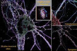
To further explore these large data sets, the research team developed a machine learning-based analysis pipeline. By analyzing 1600 features from the imaging datasets, such as protein content, distribution, or shape, the scientists discovered a previously unknown type of chemical synapse. These synapses make up only about 1% of all synapses. They would not have been detected with other imaging techniques.
With SUM-PAINT, the team provides an integrated workflow for data generation and analysis that can be used by researchers around the world. SUM-PAINT is relatively easy to use with commercially available microscopes.
"We are convinced that SUM-PAINT is not only a milestone on the way to deciphering the complexity of cell biology at the molecular level, but also a potential breakthrough in the discovery of new therapeutic approaches for neurodegenerative diseases," says Ralf Jungmann, head of the Molecular Imaging and Bionanotechnology research group at MPI of Biochemistry and holder of the Chair of Molecular Physics of Life at LMU.
By providing a detailed view of the location and interaction of a large number of proteins at the molecular level, SUM-PAINT opens up unprecedented opportunities to investigate previously hidden details of neurological disorders. In this way, the new method could contribute to a deeper understanding of the underlying mechanisms of diseases such as Parkinson's or Alzheimer's dementia.
[tb]
Dictionary of the Research Group Molecular Imaging and Bionanotechnology:
DNA-PAINT: DNA-PAINT is a high-resolution imaging technique developed by the research group of Ralf Jungmann. It involves the temporary binding of short, dye-labeled DNA strands to their target molecules. When they bind, they generate a flash of light. This allows the target molecules to be localized and imaged.
Proteome: The proteome is the totality of all proteins in a living organism, tissue, or cell at a given time. The proteome is highly dynamic and responds to the needs of the cell as well as to disease or environmental influences.
Synapse: Synapses are the points of contact between two nerve cells. They are used to transmit information in our brain.
Cytoskeleton: The cytoskeleton is made up of proteins and serves to stabilize the cell.


