Imaging Facility
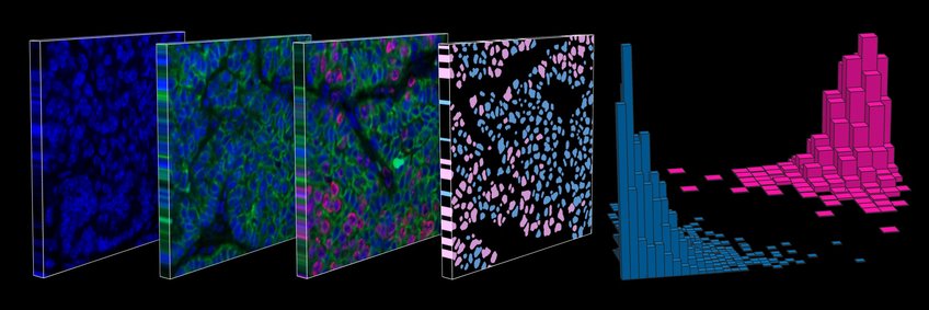
The Imaging Facility gives researchers access to sophisticated fluorescence microscopes for the observation and analysis of live or fixed organisms and cells. The scale of samples ranges from whole (small) organisms to single molecules with super-resolution microscopy. We also have high-end flow cytometers, which allow analysis and sorting of cells in many fluorescence channels at high speed. Users get personal training to use equipment and software as needed for their specific experiments, as well as support for data processing and especially quantitative image analysis.

Microscopy
Currently we have six light microscopes:
- WF1: Widefield microscope GE DeltaVision Elite, including laser module for TIRF and photo bleaching /switching)
- WF2: Widefield microscope Leica Thunder with high-sensitivity sCMOS camera, image splitter and lasers for TIRF and fast scanning for photo switching / bleaching
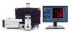
- CF1: Laser-scanning confocal microscope Zeiss LSM780, including a module for FCS and RICS
- CF2: Laser-scanning confocal microscope Leica SP8 FALCON, including White Light Laser and fully integrated FLIM module
- CF3: Laser-scanning confocal microscope Zeiss LSM980 with Airyscan 2 module for higher resolution, sensitivity and speed
- SR1: Super-resolution microscope Zeiss Elyra PS.1 with modules for SMLM (PALM, STORM, PAINT) and SIM, as well as dual-camera module with image splitter
- HCS1: Imaging robot Perkin Elmer Opera Phenix for fully automated image acquisition of many samples for hight-content screening
All systems are equipped with motorised stage, temperature control and CO2 incubation.
Flow Cytometry

For cell analysis:
- Cyto1: High-speed flow cytometer Thermofisher Attune NxT with automated plate reader
For cell sorting:
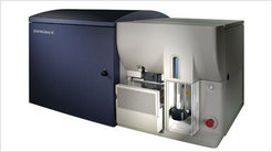
- SORT1: Fluorescence-based cell sorter BD FACS Aria III
- SORT2: Fluorescence-based cell sorter Beckman Coulter Cytoflex SRT
Alle our flow cytometers are equipped with excitation lasers from UV (375-405 nm) to far red (633-638nm) and emission filters suitable for virtually any fluorophore.

