Research
Our current work is focused onto cell division of multinucleate cells. By electric pulses we produce fused cells, in which many nuclei synchronously enter mitosis.


Patterns of PIP3 (blue), polymerized actin (red), and filamentous myosin-II in cell polarity, cell division, and particle uptake. Under certain conditions, the same components of the cell membrane and the underlying actin network can be imaged in the form of dynamic wave patterns on the substrate-attached cell surface. From Gerisch et al., BMC Cell Biol. 2011, 12:42
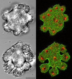
Division of multinucleate cells

Two stages of mitotic division of a multinucleate cell produced by electric pulse-induced fusion. The spindles and aster microtubules of the mitotic apparatus are labeled red, myosin-II enriched in the cleavage furrows is shown in green.
Our current work is focused onto cell division of multinucleate cells. By electric pulses we produce fused cells, in which many nuclei synchronously enter mitosis. These cells do not divide by ring-shaped cleavage furrows, as normal cells do, but by unilateral furrows. Since the molecular basis of this unusual mode of cleavage turned out to be similar to normal cytokinesis, we use the multinucleate cells as a tool to study the mechanism of mitotic cell division.
Decision making in phagocytosis
Phagocytic cells are capable of handling particles of irregular shape. The question is, how a phagocyte gathers information about the particle’s profile and its size. When a phagocyte is engaged with an oversized particle, it is uncertain whether it can completely engulf the prey. If not, it has the choice of turning uptake into release. The molecular setup of phagosomes, in particular their association with motor proteins, during this reversal of activities is under investigation.
Wave patterns in cell membrane and actin cortex

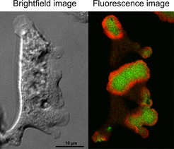
A large Dictyostelium cell produced by electric pulse-induced fusion forming actin waves. Left panel: DIC-bright field image. Right panel: Dual-color fluorescence image of the same cell showing in red actin waves and in green the phosphoinositide PIP3 at the membrane area circumscribed by the waves.
Propagating actin waves separate the cell membrane into two areas that are distinguished by their phosphoinositide composition. These dynamic patterns are associated with the recruitment of specific formins, which are molecular machines that assemble actin subunits into filaments. The dynamics of this process is studied in cooperation with the group of Jan Faix at the Medizinische Hochschule, Hannover.