Insights into the structure of a protein transport assistant

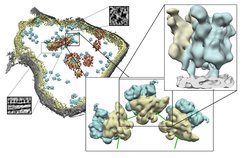
Three-dimensional structure of a mitochondrion with increasing level of details
Left: Surface representation of a mitochondrion showing the outer (grey) and the inner (yellow) membrane, mitoribosomes (blue) as well as further protein complexes (red). The small boxes show examples of the original pictures. Center: Typical assembly of mitoribosomes forming a polysome. Large (blue) and small (yellow) subunits are shown. Right: Structure of a mitoribosome (yellow and blue) bound to the inner membrane of the intact mitochondrion.
Producing a protein is a highly intricate process for the cell and involves many individual steps. Depending on the purpose for which a protein is used, there are different sites for protein production: the cytoplasm or the endoplasmic reticulum (ER). The ER is separated by a membrane from its surroundings in the cytoplasm. Even before protein synthesis is completed, the proteins produced at the ER enter via its membrane into the interior of the ER and are modified through the attachment of sugar residues concomitantly. Without these attachments, the proteins would not be able to fold properly and thus would not fulfill their functions in the cell.
Scientists of the research group “Modeling of Protein Complexes” have now described the architecture of the protein complex responsible for the transport and modification of the newly produced protein: the ER translocon. “It is located in the membrane of the ER, and this fact, together with its size and complex composition, has greatly hampered previous structural studies,” says Friedrich Förster, group leader at the MPI of Biochemistry, describing the initial situation. The structures of many subunits and their arrangement in the native ER translocon have thus far remained elusive.
It was not until cryoelectron tomography came into use that researchers could gain first insight into the architecture of the translocon. The sample is “shock frozen” to preserve its natural structure. Using an electron microscope, the scientists capture two-dimensional images of the object from different perspectives, from which they then reconstruct a three-dimensional image. Further investigations have made it possible to identify individual modules in the structure. Among them is the module that attaches the sugar residues to the newly produced protein.
“Based on this method, we will now try to determine the structure and location of other components of the ER translocon," says Förster. If the researchers know the individual structures of the ER translocon and their arrangement in the complex, they can indirectly draw conclusions about the precise functions and interactions of all components.
Original publication
Pfeffer, S., Dudek, J., Gogala, M., Schorr, S., Linxweiler, J., Lang, S., Becker, T., Beckmann, R., Zimmermann, R., Förster, F.: Structure of the mammalian oligosaccharyl-transferase complex in the native ER protein translocon. Nature Commun, January 10, 2014
DOI: 10.1038/ncomms4072 (2013).


