New Force Sensing Method Reveals How Cells Sense Tissue Stiffness

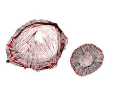
The cell adhesion protein talin (shown in red) bears mechanical forces of seven to ten piconewton und mediates the engagement of cell adhesions to the actin cytoskeleton (shown in grey). This talin linkage allows cells to sense the stiffness of their environment so that cell adhesions become reinforced on stiff substrates (left cell). Impairing the mechanical talin linkage leads to a loss of cellular rigidity sensing (right cell).
To measure the exerted molecular forces, Carsten Grashoff and his team have engineered two novel fluorescent biosensors that change their color in response to piconewton forces. Genetic insertion of these biosensors into the protein of interest allows the microscopic evaluation of molecular tension in living cells. About five years ago, Grashoff presented the first tension sensor prototype, which has been used in many laboratories worldwide, but he is confident that the new technique will be applied by many research teams as well. “The new probes enable us to resolve differences in mechanical tension with much higher precision,” said study leader Grashoff whose research group is supported by the Max and Ingeburg Herz Foundation.
By applying the method to the cell adhesion protein talin, the researchers at the Max Planck Institute discovered the central mechanism that allows cells to feel their mechanical environment. “Talin carries mechanical force of about seven to ten piconewton during cell adhesion,” Grashoff explained. “We expected that talin is involved in the force sensing process but we had no idea how important it really is. Cells in which talin is not able to form mechanical linkages can no longer distinguish whether they are on a rigid or a soft surface.”
The researchers also found that a second talin protein, called talin-2, can be used by cells to adapt to very soft environments. Since talins are present in all cells of our body, the researchers believe that they have found a general mechanism cells use to measure the extracellular rigidity. The new findings were published in the journal Nature Cell Biology.
Original publication:
K. Austen, P. Ringer, A. Mehlich, A. Chrostek-Grashoff, C. Kluger, C. Klingner, B. Sabass, R. Zent, M. Rief, C. Grashoff: Extracellular rigidity sensing by talin isoform-specific mechanical linkages. Nature Cell Biology, November 2015
DOI: 10.1038/ncb3268


